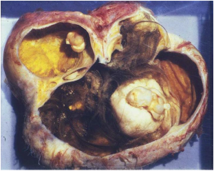
These cysts contain many types of cells. They may be filled with hair, teeth, and other tissues that become part of the cyst. They can become large and cause pain.
Based on research, the best way to get rid of dermoid ovarian cysts fast is by using this treatment.
Dermoid cysts, also known as benign or mature cystic teratomas(MCT), are composed of mature tissues, which can contain elements of all three germ cell layers. They have been reported in patients aged 1–91 years. They are the most common ovarian neoplasm found in adolescence and during pregnancy. While this tumor is usually asymptomatic, 47% of patients complain of abdominal pain.
Although ovarian dermoid rupture and Wstula formation involving the bladder, rectum, and intestine are well documented in malignant transformation in the gynecologic literature, Wstulation in benign cystic teratomas has not been commonly reported and is thus not well recognized by physicians.
Complications associated with dermoid cysts include torsion, rupture, infection, malignant transformation, fistula formation, and hemolytic anemia. Since the wall of denmoid cysts is thick, rupture is rare and therefore potentially misinterpreted.
Possible explanations for rupture on perforation include partial torsion of the tumor, resulting in circulatory compromise and subsequent tumor wall necrosis; infection; malignant transformation; and mechanical factons. Although this complication is rare, it is potentially devastating; the most threatening sequelae include acute peritonitis and hemorrhagic shock. Ovarian cyst treatment.
Acute rupture of a dermoid cyst with acute peritonitis is usually associated with pregnancy and labor. Intnaabdominab spillage of tumor contents results in severe chemical pentonitis, granubomatous reaction, and formation of dense adhesions. Adhesions of tumor to adjacent structures and the inflammatory reaction may result in a fistula and drainage into another abdominal organ. Numerous reports document spontaneous ruptune of denmoid cysts into the bladden, small bowel, rectum, sigmoid cobon, and vagina and perforation of the anterior abdominal wall.
In contrast to patients with acute cystic rupture and resultant penitonitis, patients with chronic involvement may present with progressive distention, anorexia, nausea, vomiting, or diarrhea. If rupture occurs into a hollow viscus, the situation is generally not suspected until hair, teeth, on sebaceous material are passed through the orifice of the natural organ. Since this potentially serious condition is rarely diagnosed correctly in a clinical evaluation, radiographic analysis is critical.


 RSS Feed
